resources


resources

Sunday, February 15, 2009
Endocardial cushion defect (ECD) is an abnormal heart condition in which there is no separation between the chambers of the heart. Essentially, the middle part of the heart is missing. It is a congenital heart disease, which means it is present from birth.
Causes
Endocardial cushion defect occurs while a baby is still growing in the womb. The endocardial cushions are two areas of thickening that eventually develop into the wall (septum) that separates the four chambers of the heart. They also form the mitral and tricuspid valves.
The lack of separation between the two sides of the heart causes several problems:
•Increased blood pressure in the lungs. In persons with this condition, blood flows through the abnormal openings from the left to the right side of the heart, then to the lungs. The increased blood flow into the lungs leads to a rise in blood pressure in the lungs.
•Lung irritation and inflammation. Increased blood flow into the lungs causes irritation and swelling.
•Heart failure. Because the heart has to pump more blood to the lungs, it has to work much harder than normal. The heart may enlarge and weaken.
•Lack of oxygen. As the blood pressure increases in the lungs, blood flow starts to move from the right side of the heart to the left. The heart starts pumping oxygen-poor blood throughout the body, and will have to work harder at it. The body will not get enough oxygen.
There are two types of ECD:
•Complete ECD: A complete ECD involves an atrial septal defect (ASD) and a ventricular septal defect (VSD). Persons with a complete ECD have only one large heart valve (common AV valve) instead of two distinct valves (mitral and tricuspid).
•Partial (or incomplete) ECD: Only an ASD is present. There are two distinct valves, but one of them (the mitral valve) is often abnormal with an opening ("cleft") in it, letting blood leak between the two left chambers of the heart.
ECD is strongly associated with Down syndrome. Several gene changes are also connected to ECD. However, the exact cause of ECD is unknown.
ECD may be associated with other congenital heart defects such as:
•Double outlet right ventricle
•Single ventricle
•Transposition of the great vessels
•Tetralogy of Fallot
Symptoms
Symptoms of ECD may include:
•Baby tires easily
•Bluish skin color (the lips may also be blue)
•Failure to gain weight and grow
•Frequent pneumonia
•Lack of appetite
•Pale skin (pallor)
•Rapid breathing
•Rapid heartbeat
•Sweating
•Swollen legs or abdomen (rare in children)
•Trouble breathing, especially during feeding
Exams and Tests
Signs of ECD may include:
•An abnormal electrocardiogram (ECG)
•An enlarged heart
•Heart murmur
•High blood pressure in the lungs
Children with partial ECD, who have only a small VSD and normal valves, may not have signs or symptoms of the disorder during childhood.
Tests to diagnose ECD include:
•Ultrasound of the heart (echocardiogram) to see blood flow
•An electrocardiogram (ECG), which measures the electrical activity in the heart
•Chest x-ray, which may show an enlarged heart
•Magnetic resonance imaging (MRI) of the heart, which provides a detailed image of the heart through the use of powerful magnets
•Cardiac catheterization (in some cases), a procedure in which a thin tube (catheter) is placed into the heart to see blood flow and take accurate measurements of blood pressure and oxygen levels
Treatment
Surgery is needed to close the holes between the heart chambers, and build new tricuspid and mitral valves. The timing of the surgery depends on your child's condition and the severity of the ECD, but it can usually be done when the baby is about 3 months old. Correcting an ECD may require more than one surgery.
Your doctor may prescribe medication before surgery if the ECD has made your baby very sick. The medicines will help the child gain weight and strength before surgery. Medications may include:
•Diuretics (water pills)
•Drugs that make the heart contract more forcefully (inotropic agents), such as digoxin
Surgery for a complete ECD should be done as early in the baby's first year of life as possible, before irreversible lung damage occurs. Babies with Down syndrome tend to develop lung disease earlier, and therefore early surgery is very important for these babies.
Outlook (Prognosis)
How well your baby does depends on the severity of the ECD, the child's overall health, and whether lung disease has already developed. Many children live normal, active lives after the ECD is corrected.
Possible Complications
Complications from ECD may include:
•Congestive heart failure
•Death
•Eisenmenger syndrome
•High blood pressure in the lungs
•Irreversible damage to the lungs
Certain complications of ECD surgery may not appear until the child is an adult. These include heart rhythm problems and a leaky mitral valve.
All children with congenital heart disease should take antibiotics before dental treatment. This helps prevent complications related to heart infections.
When to Contact a Medical Professional
Call your health care provider if your child seems to tire easily, has trouble breathing, or has bluish skin or lips. You should also consult your health care provider if your baby is not growing or gaining weight.
Prevention
ECD is associated with several genetic abnormalities. Couples with a family history of ECD may wish to seek genetic counseling before becoming pregnant.
Alternative Names
Atrioventricular (AV) canal defect; Atrioventricular septal defect; AVSD
References
Park MK. Pediatric Cardiology for Practitioners, 5th ed. Philadelphia, Pa: Mosby Elsevier; 2008:181-189:chap 12.
Townsend Jr. CM, Beauchamp RD, Evers BM, et al. Sabiston Textbook of Surgery, 18th ed. Philadelphia, Pa: Saunders Elsevier; 2008:1760-1762.

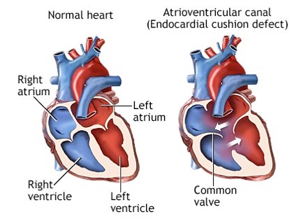
Atrioventricular Canal (Endocardial Cushion Defect)